Clinical Center Orthopectus
World Reference in Pectus Treatment
Studying and providing Pectus care since 1977.
The world’s largest experience in the conservative treatment of thoracic deformities.
Our Specialties
Chest
Backbone
Pediatric Orthopedics
Foot and Ankle
Orthotics and Prosthetics
Rheumatology
Sports Medicine
Shoulder, Elbow and Hand
Physiotherapy
Our Specialties
Chest
Backbone
Pediatric Orthopedics
Foot and Ankle
Orthotics and Prosthetics
Rheumatology
Sports Medicine
Shoulder, Elbow and Hand
Physiotherapy
Our History
Studying and providing Pectus care since 1977.
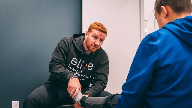
testimonials
What our patients say about Orthopectus:


News and Information
In summary pectus should always be treated with braces and specific exercises (Haje’s method) as the first treatment choice for pectus carinatum …
The deformities of the anterior chest wall are known as pectus carinatum or “pigeon breast” and pectus excavatum or “shoemaker chest”. They are very common, affecting …
In 2016, Dr. Davi Haje, Dr. Moacir Silva and Dr. Sydney Haje (in memoriam) will launch a new book’s chapter on pectus …
In June 2016, Dr Davi Haje delivered an important lecture about the Haje’s method of treating pectus in the next CWIG meeting, …
Download the definitive manual on Pectus now
Have your questions answered by the world’s largest reference on the subject